Shang Wang Advances Biomedical Imaging with Open Source 4D Tools
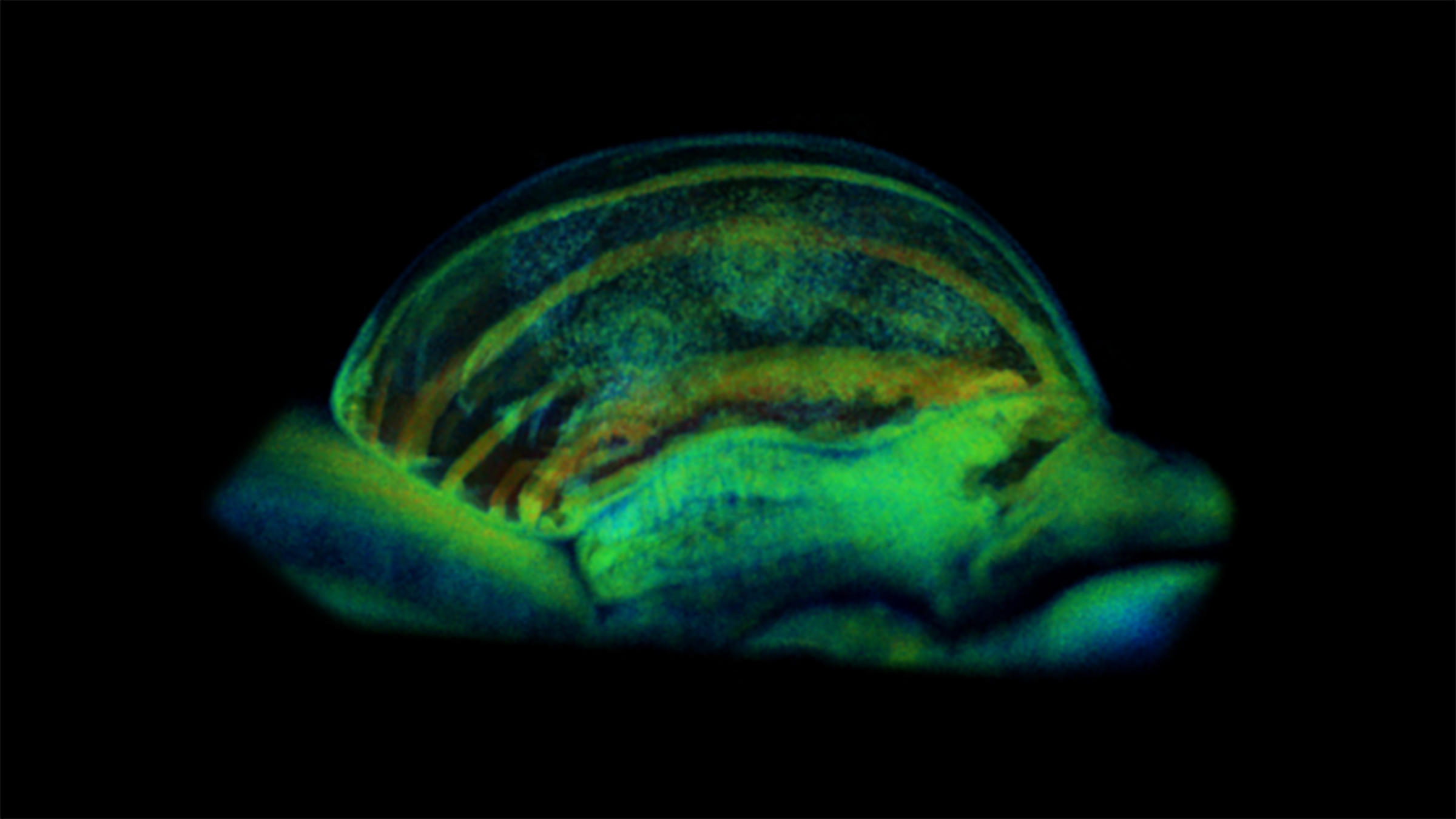
Understanding how fundamental biological processes work is essential to biomedical research. However, observing cells and tissues as they function in three dimensions within their natural environment requires a combination of scale, speed and contrast that traditional imaging techniques have not been able to achieve.
Shang Wang, associate professor, Department of Biomedical Engineering, is working to improve this outlook. By developing innovative optical imaging and computational tools, his research helps scientists visualize and analyze biological processes as they happen — not only in 3D, but also across time. This 4D view offers a more complete picture of how biological systems actually work.
“Mechanical factors, such as force and strain, are critically important cues regulating physiology at the cell and tissue levels,” Wang explained. “Thus, biomechanical process, such as how a biological system applies mechanics to achieve its function, is one of our primary targets.”
Using light-wave technology known as optical coherence tomography (OCT), Wang and his team have developed high-resolution imaging methods that capture and integrate these kinds of biological information. The resulting images reveal detailed tissue structures and functions, as well as their changes.
To turn these massive imaging datasets into useful insights, the team also built user-friendly, open-source software tools: Open Chrono-Morph Viewer (OCMV) and an interactive visualization technique called the clipping spline.
A previous National Institutes of Health (NIH) Maximizing Investigators' Research Award (MIRA) is supporting the development of these methods and tools.
“Medical imaging allows us to see inside the body without having to ‘open up’ the patient,” said Jennifer Kang-Mieler, chair of the Department of Biomedical Engineering and George Meade Bond Professor. “Shang’s work takes that concept to another level. Integrating imaging technology, computation and biology gives us a much richer picture than a traditional static image and lets us study processes that were previously difficult to observe.”
Getting to the heart of the matter
The innovations grew out of embryonic heart development studies in Wang’s laboratory. When the researchers found that existing tools could not help them fully visualize and interpret their 4D OCT data, they simply built their own.
OCMV is a standalone software program for analyzing 3D bioimages over time. One of its distinctive features is the clipping spline tool, which lets users cut away portions of a 3D along a flexible, curved path to examine structures inside it. This work was recognized in 2025 as a Biomedical Optics Express Editors’ Pick and cover feature.

With OCMV, researchers can directly visualize 4D data from an embryonic heart, view multiple parts of the developing organ at once and analyze its biomechanics. As 4D imaging capabilities advance, the tool could support research far beyond cardiac development.
For example, Wang’s team is also using the technology to study how the fallopian tube, or oviduct, moves preimplantation embryos toward the uterus. They hope to reveal insights into the causes of oviduct-related infertility and tubal ectopic pregnancies.
“Through 4D imaging, visualization and analysis with a mouse model, we uncovered how the muscular activity transports the embryo,” he said. “This creates a new understanding in relation to the function of the oviduct in pregnancy.”
Their work on heart development and embryo transport is already demonstrating how studying biological processes in action can pave the way for transformative findings.
“Cells move, tissues develop and remodel and physiological processes change over time, but we often study these processes by looking at individual snapshots,” Kang-Mieler said. “Shang’s work allows us to observe these processes as they are happening, in three dimensions and at very high resolution, without disrupting the biological system. When you combine that imaging with computational tools that can extract and quantify information from large, complex datasets, it opens up new ways to understand both normal biology and disease.”
Building a community around 4D imaging for better health outcomes
For Wang and his team, making OCMV open source was an easy decision.
“We hope other researchers can use these tools to address their needs in handling, visualizing and analyzing 4D image data,” Wang said. “It allows them to build on what we have developed to achieve further advancements more efficiently. Ultimately, we hope this will be a community-driven platform for bioimage informatics, accelerating discoveries in biomedicine.”
Ultimately, that is the power of biomedical technology development: improving how researchers measure and understand biological processes, then using that knowledge to advance human health.
“Innovations in imaging drive biomedical discoveries,” said Wang, who will be honored as one of two Distinguished Young Engineering Alumni at the University of Houston Cullen College of Engineering’s 2026 Cullen Gala. “I hope the bioimaging tools we develop will have a broad impact by enabling many new studies in biomedicine. I also hope our fundamental biological findings will lead to new strategies and approaches in medicine.”
Learn more about academic programs and research in the Department of Biomedical Engineering: