Seeing Stroke Risk in a New Light: Wearable Laser Tech to Read the Brain
When Biomedical Engineering Assistant Professor Simon Mahler describes his research, he starts with a simple image: a red laser beam passing through frosted glass. The grainy “snow” that appears, also known as laser speckle, is usually something optical engineers try to eliminate. Mahler learned to suppress it in nanoseconds during his Ph.D. But then he flipped the idea on its head. What if those shifting speckles could become a signal to tap into—not noise to eliminate—for measuring dynamic, real-time blood flow in the human brain?
From France to physics, optics to blood flow
 Mahler grew up in Marseille, France, and earned a bachelor’s degree in physics at Aix-Marseille University. He then completed a dual master’s in fundamental physics and complex systems at Universités Paris-Sud and Paris-Saclay and during this time became drawn to working with laser light.
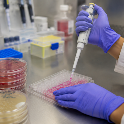
“I was feeling more attracted by optics, especially lasers,” he explained, “so I specialised in optical measurement at the end of my master.” During his Ph.D. at the Weizmann Institute of Science in Israel, he built degenerate laser cavities and explored dynamic speckle suppression, originally aiming to improve laser beam uniformity for coupling lasers. But the turning point came when he realized, “Instead of suppressing the speckles, we can use them.” The grainy laser speckle effects once considered a nuisance could become a rich source of information about motion and flow. That realization led to the familiarization of Mahler with laser speckle imaging and the development of compact optical systems capable of mapping vascular flow in embryos such as chicken eggs—Laser Speckle Contrast Imaging (LSCI)—and, eventually, in humans—“Speckle Contrast Optical Spectroscopy” (SCOS).
A breakthrough idea in optics
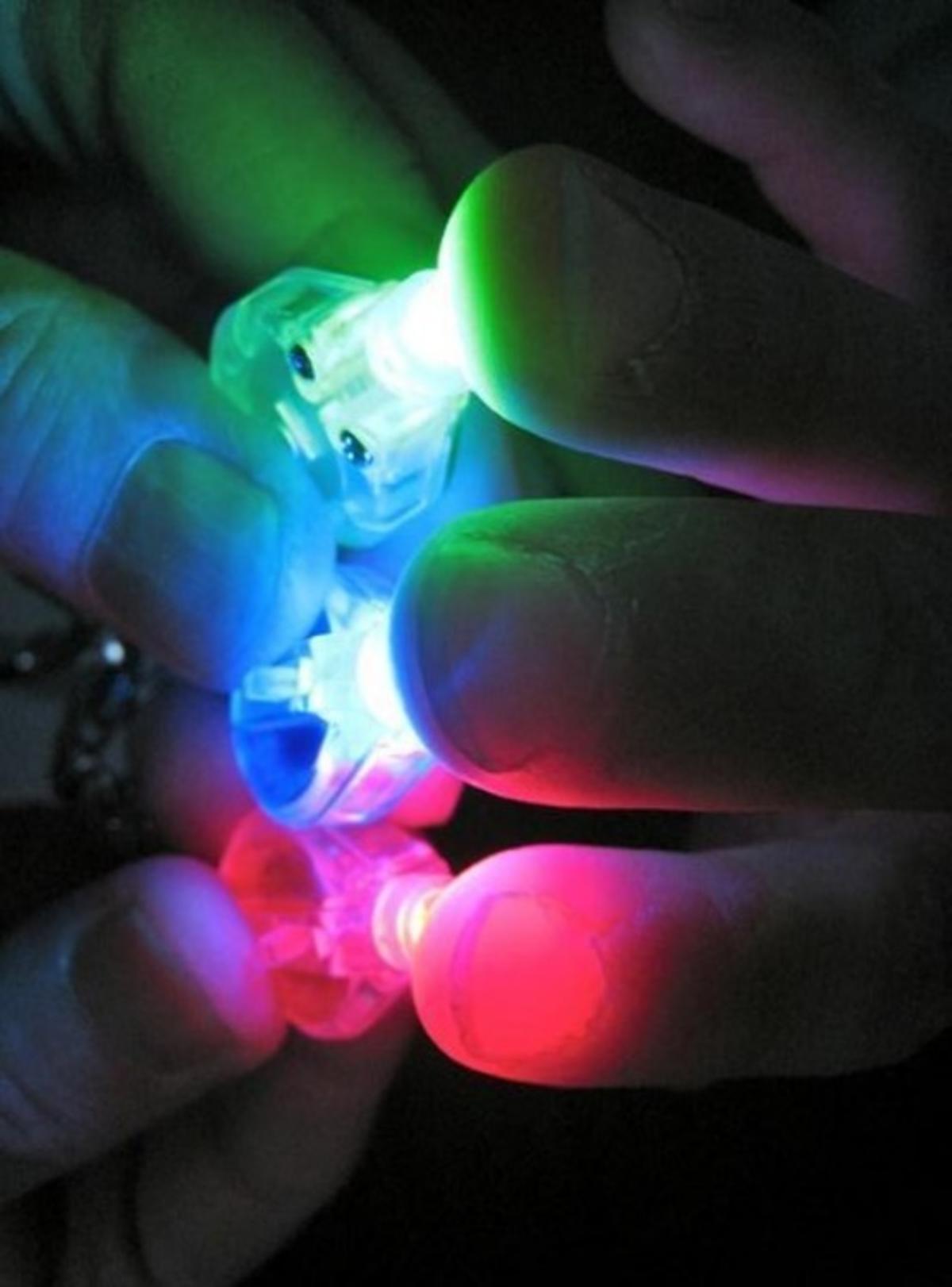
 At the heart of Mahler’s work is near-infrared light, which penetrates skin and skull deeper than visible wavelengths. Military ops and specialized rescue teams have used infrared technology for decades for its body heat-detecting capabilities in the dark.
In recent years, regular people around the world have been introduced to near- and far-infrared light in the form of infrared saunas, which have been proven to give increased detoxification for a shorter duration at lower temperatures than a traditional coal-burning sauna. The same infrared light that can help us locate people and promote wellness can also peer into our skulls.
When this light scatters through moving red blood cells in the brain, it creates a changing speckle pattern. By capturing those fluctuations at high speed and measuring “speckle contrast,” Mahler and his team can infer how quickly blood is flowing and how vessels are reacting. Using a simple rotating diffuser as a lab model, he showed how motion reduces speckle contrast and creates clearer, more uniform speckle frames.
“If nothing moves, the speckle contrast is high. When blood is moving fast, the pattern smooths out.”
A five-minute “brain stress test”
In a landmark human study published in Biomedical Optics Express, Mahler and colleagues proved using a head mounted SCOS device that the principle holds: faster-moving cerebral blood produces smoother speckles; slower or impaired flow produces higher contrast. Through a controlled breath-holding task, they observed how blood flow and blood volume changed in real time. The key metric: a flow-to-volume ratio. Participants at higher risk of stroke exhibited markedly higher ratios, meaning their vessels could not dilate effectively under increased demand, so blood flow increased but blood volume did not.
“Seeing that flow-to-volume split was the ‘aha’ moment,” Mahler says. “It’s a physiological marker you can measure without an MRI or contrast agents.”
“That’s why I chose a biomedical engineering department, so I could combine physics, optics and medicine."
Replacing unreliable risk predictor with hard data
Stroke remains a leading cause of disability and death worldwide, and Mahler’s headset could eventually become the clinical standard for a noninvasive stroke-risk screen that could be done quickly and at low cost–opening doors to preventive care rather than crisis response. Today, clinicians estimate stroke risk largely from questionnaires (age, blood pressure, smoking and alcohol history, etc.) and lab values, rather than direct measurements of vessel health.
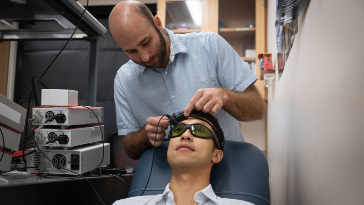 “Instead of using a questionnaire, we put our device on participants and ask them to hold their breath. We then measure their brain’s blood-flow response,” Mahler explained. “Breath-holding mimics a stress test and we can see how the brain performs under a sudden lack of oxygen.”
The result: a physiologic signature rather than a paper estimate. Mahler’s work adds a direct, brain-based physiological readout, addressing a gap often noted in preventive neurology. While most people know, in theory, that their lifestyle contributes to vascular risk, seeing those effects visualized through hard data could serve as a powerful wake-up call.
“It’s always better to see a problem coming than to deal with it after,” he emphasized.
Ultimately, his goal is to shift the paradigm from reactive stroke care to proactive brain-health monitoring. If this technology reveals unexpectedly high stroke-risk patterns in specific populations—or shows large cross-country differences—it could influence public-health policy as well.
Mapping traumatic brain injury, too
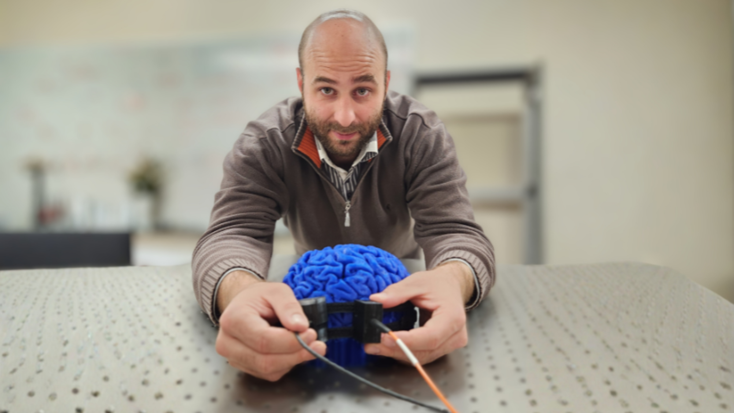
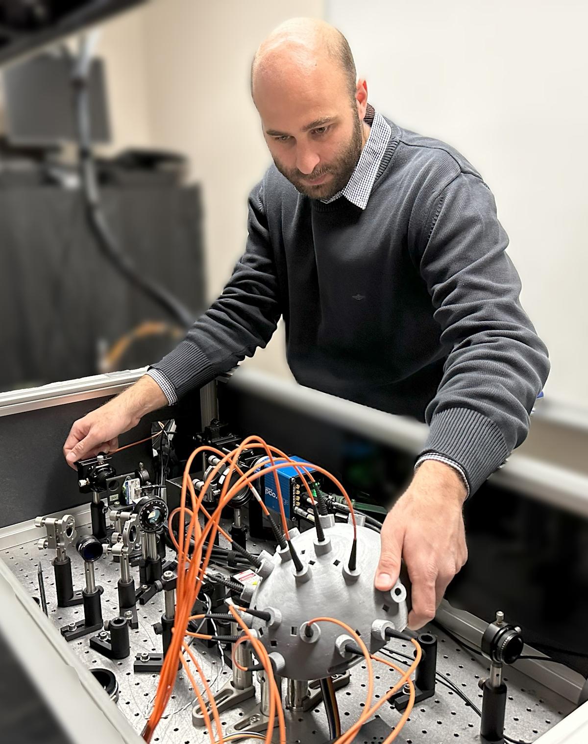
 Mahler’s group also built a six-channel version of the system that samples blood-flow dynamics from multiple sites across the head.
In preliminary tests, channels overlying a brain-injury site showed distinctly abnormal waveforms compared with healthy regions—hinting at potential for bedside monitoring of traumatic brain injury or aneurysm patients.
His multi-channel wearable could one day allow clinicians and individuals to monitor cerebrovascular compliance, track recovery after brain injury, and screen for early signs of vessel dysfunction. The device’s advantages: faster, cheaper, and more accessible than MRI or CT-based perfusion scans.
What’s next: multi-disciplinary research for a far-reaching revolution
Mahler is now establishing his speckle imaging lab at Stevens. He has two primary research streams: one follows on cardiovascular and cerebrovascular development in chick embryos using speckle devices; the other scales the technology for clinical use in stroke risk assessment and brain-injury monitoring. His journey—from physics to optics to biomedical engineering—reflects a broader ethos: bringing tools from one domain into entirely new applications in another.
“That’s why I chose a biomedical engineering department, so I could combine physics, optics and medicine," he said.
His collaborators span laser physicists, neurosurgeons, biologists, and engineers. This multidisciplinary mix is what makes the innovation possible. The research may start with speckle patterns and lasers, but the implications reach into prevention, diagnostics and ultimately patient care.
As the device evolves and enters larger clinical trials, it may well reshape how we think about brain health. For Mahler and his team, the vision is clear: a world in which a simple optical scan warns you of vessel stress before it becomes life-or-death. “A laser and a camera,” he said—offering a window into the brain that until now didn’t exist.
Learn more about academic programs and research in the Department of Biomedical Engineering: