Changing How We See Cancer: Interdisciplinary Stevens Team Receives NCI Grant of $450,129 to Image Ovarian Cancer Metastasis
Stevens professors Marcin Iwanicki and Shang Wang received a National Cancer Institute grant for imaging of ovarian cancer metastasis after chemotherapy
According to the American Cancer Society, this year alone, more than 21,000 women in the United States will find out they have ovarian cancer—and another 13,000 women will die from the disease. These depressing statistics are due to a lack of early detection strategies and the development of chemotherapy resistance.
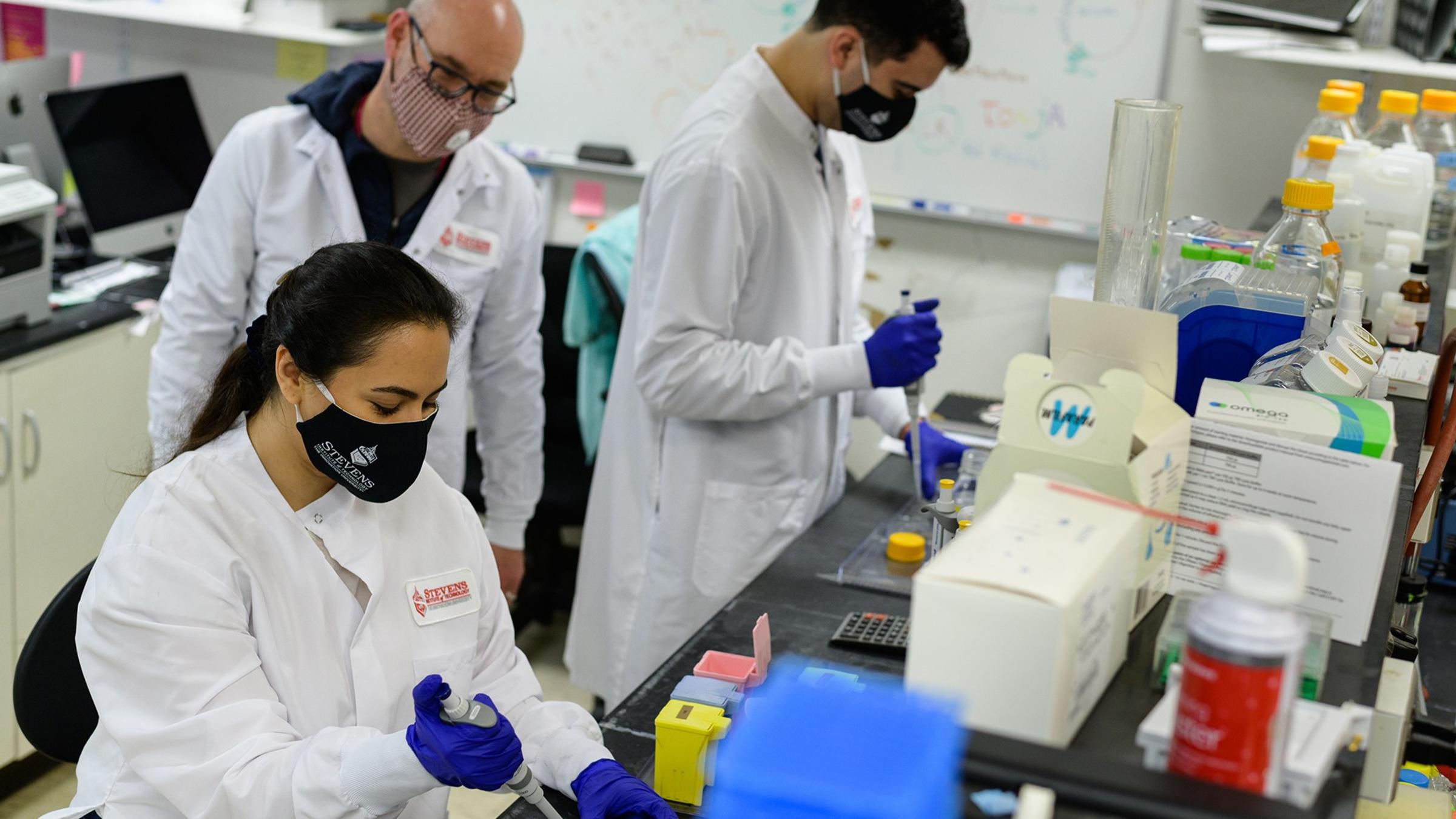
Two researchers at Stevens Institute of Technology—Marcin Iwanicki, assistant professor in the Department of Chemistry and Chemical Biology, and Shang Wang, assistant professor in the Department of Biomedical Engineering—are bringing their areas of expertise together to change how researchers view ovarian cancer. Literally. The team received a National Cancer Institute (NCI) grant of $450,129 titled “High-Resolution Dynamic Imaging of Ovarian Cancer Metastasis Post-Chemotherapy,” which will develop high-resolution optical imaging approaches to capture post-chemotherapy ovarian cancer metastasis.
Metastasis after chemo
For patients with ovarian cancer, chemotherapy can be a double-edged sword: it may kill the targeted tumors but then those tumors might come back after treatment. “And when they come back, they're even more aggressive—meaning more metastasized—and that's what ultimately causes a patient's death,” explained Iwanicki.
Because of the location of the ovaries and the fallopian tube in the body, metastasizing tumor cells can readily detach and spread through the peritoneal cavity—and patients can only take chemotherapy so many times.
Iwanicki has been studying this problem for 12 years and has had success showing this post-chemo metastasis on 3D tissue cultures called organoids. Using those models, his team can see tumors spread after chemotherapy recovery. “It's a nice indication of this chemotherapeutic effect on tumor metastasis,” said Iwanicki. “But, in the scientific community, you have to really examine this using additional approaches.”
That’s where Wang comes in. He has been working on high-resolution imaging techniques with the mouse model, including the development of optical imaging approaches to study the function of fallopian tube. “We are interested in developing new imaging methods to answer questions in developmental and reproductive biology,” explained Wang. “And such work has led to the capability to image the tissue and cell activities in the reproductive organs.”
A new way to image cancer
Wang’s imaging research is based on a technique called optical coherence tomography, which is similar to ultrasound imaging but with a higher resolution for seeing structures in more detail. In bioimaging, there is a tradeoff between resolution and imaging depths, so ultrasound can get deeper images but without much detail. Optical coherence tomography does the opposite, giving much better detail but at lower depths.
This advantage of higher resolution makes it possible to measure detailed features of the tissue structure, which is critical for further understanding the tissue and cell behaviors. The imaging approach also acquires 3D images at a high speed, allowing researchers to see fast changes inside the tissue.
At Wang’s interview for a faculty position at Stevens in 2019, he and Iwanicki, who has been at Stevens since early 2018, discussed how Wang’s imaging approach could be used to study the metastasis of tumors by visualizing and quantifying their activities.
“After talking with Marcin during my interview, it was a great thought that we can collaborate on this work to address some important questions and get a much better understanding of ovarian cancer from a dynamic view with a better resolution,” said Wang. “Once you understand the disease better, you'll be able to manage it better.”
The two began writing grants together, hoping to combine their research, and in December, the professors received two years’ of NCI funding for the project. “The review panel really picked up on this beautiful merge of bioengineering and chemical biology to address this really important health problem that drugs that treat cancer actually also promote cancer,” noted Iwanicki.
Changing how we see cancer
Ultimately, the team hopes this advanced imaging will shed light on ovarian cancer metastasis. If they can confirm that chemotherapy contributes to tumor spread after treatment, the scientific community can focus on the mechanisms that drive metastasis and perhaps come up with drugs that patients could take to minimize it. That would move the needle toward Iwanicki’s ultimate hope that someday late-stage cancer will become a manageable, chronic condition like high cholesterol rather than a non-treatable disease.
For Iwanicki and Wang, the key to successful research is teamwork. “Stevens provides this very nice environment for people to work together, and the way to increase the rate of great discoveries is to always work with someone,” said Iwanicki. “I’m a cancer biologist, and Shang is a biomedical engineer—very different fields—but I've learned a lot from him already. And this is before we actually started working, so there’s lots more to come.”
Learn more about chemistry and chemical biology at Stevens:
Chemistry and Chemical Biology Department
Chemistry and Chemical Biology Undergraduate Programs
Chemistry and Chemical Biology Graduate Programs
Learn more about biomedical engineering at Stevens:
Learn more about research in the Department of Biomedical Engineering →