From Avocados to Arteries and Cancers, This AI Sees More Clearly
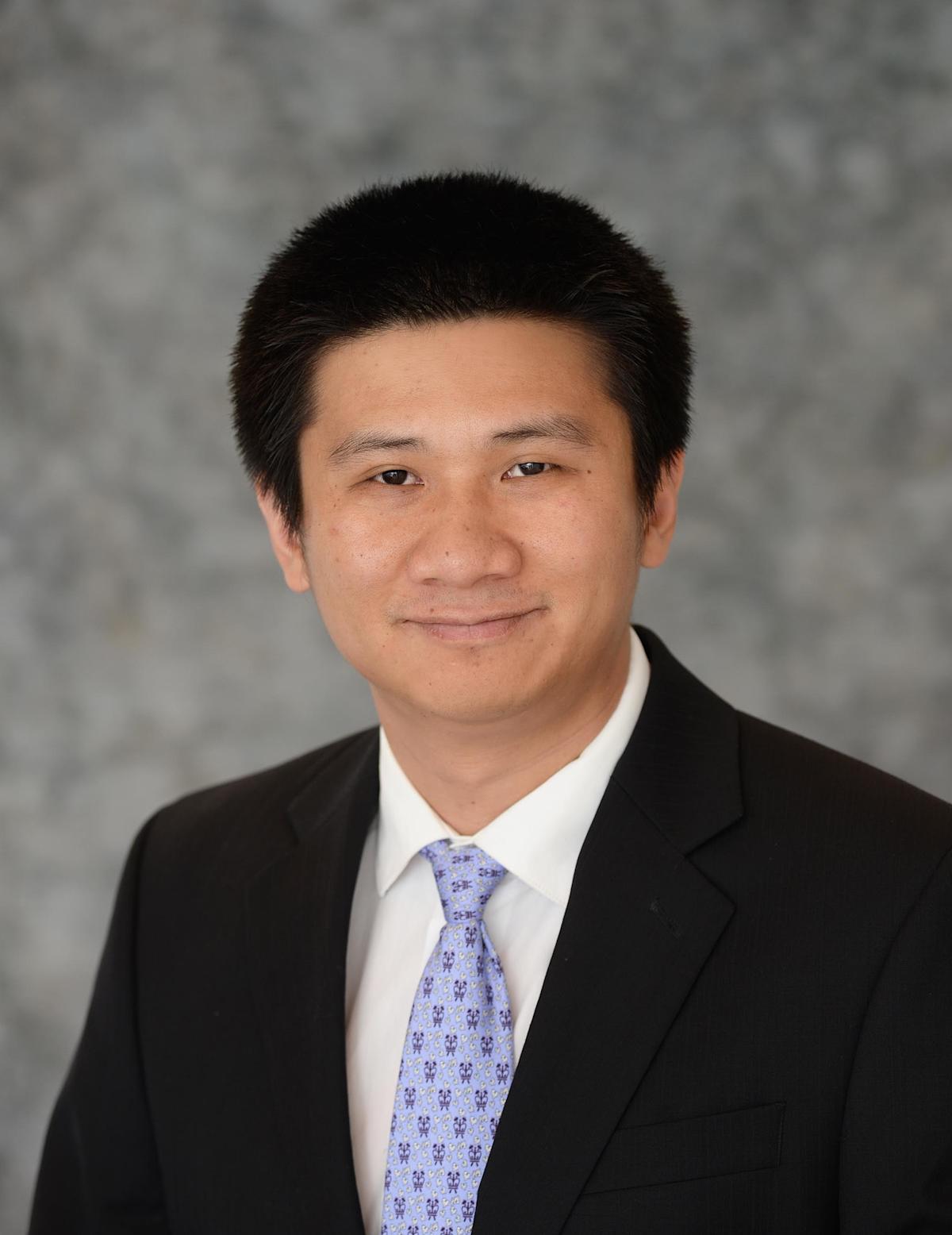
Stevens professor Yu Gan develops algorithms to improve medical diagnosis, food inspection and more
For centuries, physicians relied upon their own eyes, medical training and experience to make diagnoses and order tests.
A host of rapidly evolving medical-imaging technologies has since helped improve the odds of catching dangerous diseases early, before they become deadly — but good tools to process and analyze that data beyond what doctors’ eyes see in medical images had lagged behind.
Now the explosive rise of artificial intelligence technologies has changed the game, and Stevens biomedical engineering professor Yu Gan is using the powerful tools of AI to sharpen images, develop new health insights — and maybe even help us pick out fresher food at the grocery store.
“Better medical imaging and image processing is the future of health,” says Gan, who directs the university’s Intelligent Imaging and Image Processing (I3P) Lab and is supported by the National Institutes of Health, National Science Foundation (NSF), National Institute of Food and Agriculture, NVIDIA, Burroughs Wellcome and the New Jersey Health Foundation, among others. “That’s clear.”
Spotting hidden heart attacks, untangling cancers
As a Ph.D. student at Columbia University, Gan believed he was headed for a career in electrical engineering until he began working with the then-new tools of AI and machine learning.
 “I became fascinated by the advances in data science and machine learning. I realized right away that we could use algorithms to enhance images, unveil clinical information and improve people’s health and their quality of life,” he recalls. “That quickly became my Ph.D. dissertation and then my career path.”
Gan pursued two lines of inquiry at Columbia: the enhancement of cervical images, in an effort to prevent preterm births, and studies of images of cardiac muscle to try to early-identify heart disease. Later, after completing his doctorate and becoming a faculty member at the University of Alabama, he dialed in on internal imaging of the heart’s arteries — now the main (but not only) focus of his work.
“Atherosclerosis — hardening or blocked heart arteries — contributes to millions of deaths annually,” explains Gan. “Angiograms and other imaging tools can give doctors a visual sense of whether arteries are becoming calcified; but deep-learning methods can obtain even more accurate 3D internal views of the heart’s chambers and blood vessels, and in real time.”
After joining Stevens in the fall of 2022, Gan quickly made a mark, receiving a prestigious five-year $600,000 NSF CAREER award the following spring to support and further the work. He will use it to continue developing algorithms that produce quicker, sharper, lower-cost analyses of the images produced by optical coherence tomography, histological microscopy, MRI and other medical imaging techniques.
“In the coronary project we are looking for plaques, seeing if we can identify them in images even when they appear indistinct or ‘fuzzy’ to a human viewer,” he explains. “We want to diagnose heart disease earlier, before it strikes as a heart attack.”
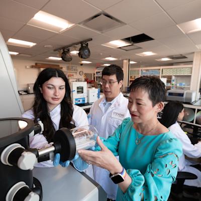
To develop his AI, Gan works closely with pathologists and cardiologists at the University of Alabama’s medical school, who supplied Gan’s lab with samples and analysis of both healthy and diseased coronary tissue.
Inside that lab, housed in Stevens’ McLean Hall, two digital imaging systems — one low-quality, one very high-quality — snap images of the tissue samples. Thousands of those images are fed as datasets into a Lambda GPU workstation across the lab, where his graduate-student team sets to work designing and training ever-better algorithms that can accurately pick out likely clogging heart arteries from clean ones.
"Eventually we will use this technology in in vivo studies with live patients as they are having intravascular imaging in medical settings,” he continues. “Our software will bring out disease patterns that were not visible at first so that physicians can see and then treat them.”
In another project, Gan’s group is investigating the use of AI to facilitate cancer detection in medical images. Working with the German Cancer Research Center (DKFZ) — Germany’s national cancer research institute — Gan’s team investigates the basic mechanisms of cancer, right down to the cellular level.
“Scientists have a lot of medical data on cancer now, but we are all still working to solve the big-data issues,” says Gan. “In our lab, we process confocal image data from tumors and try to extract the actual dynamics of cells.”
The effort, initially privately funded, is currently seeking additional support; meanwhile, his student team is helping build new software that will enable DKFZ to analyze libraries of images of tumor cells.
Fresher food: A screen-shot away?
Gan also received $300,000 in recent new funding from the U.S. Department of Agriculture for a different sort of imaging project, but one no less relevant to public health: food evaluation.
Can we one day point a smartphone packed with Stevens AI at produce in the supermarket aisle and instantly tell if it’s ripening, perfectly ripe and ready to eat... or gone to pasture? Can we tell if it contains a stone or a tiny shard of metal the eye can’t see?
“That’s the goal,” confirms Gan. “Right now we are working on training our algorithms on food images in order to teach them to accurately evaluate the freshness of food and other characteristics. Safe food, after all, is also a health issue.”
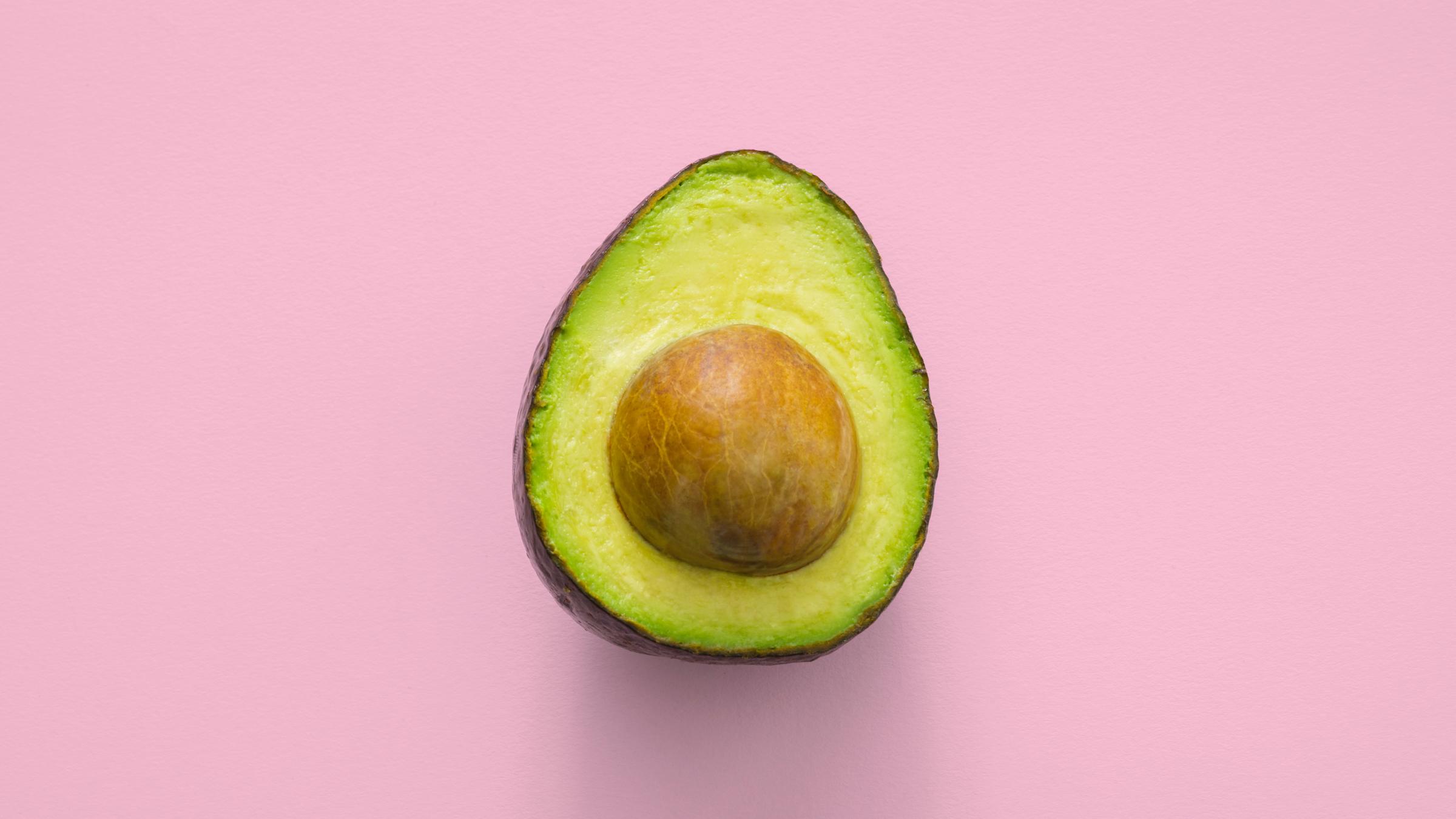
His team works with microwave images of foods such as eggs, avocados and bananas; eventually the group will move on to capturing and analyzing millimeter-wave images, a technology that has also been studied at Stevens for its potential to help diagnose skin cancer.
“We are trying to build a millimeter-wave imaging system that works at the same frequency as a 5G smartphone,” Gan says. “That’s the first step toward building an application that anyone can use.”
He's working on other problems in AI, too, including developing systems that learn from limited sample resources and ones that correct mistakes produced when training deep-learning algorithms.
“This is very important for biomedical study,” says Gan. “If you train systems with the wrong information, you may end up treating a patient incorrectly, missing a disease or treating the wrong disease. You could literally cost someone a life.”
And that’s not all. Gan hopes to design novel image-sharpening tools that could potentially detect early stages of Alzheimer’s disease from brain MRI images.
He's keeping a foot in the classroom, too, teaching a Stevens graduate-level course every year and designing innovative new coursework.
“If you are a biomedical engineering student who wants to do machine learning, there are just not very many resources, books or courses yet focused specifically on the discipline.”
So he created one: Machine Learning in Biomedical Engineering, a new graduate-level class teaching the use of AI for biomedical engineering applications plus a bit of tissue engineering and biomechanics coursework mixed in.
Gan hopes his research, in combination with his other educational activities, will help Stevens develop the next generations of students and young technologists who will continue to analyze medical data and build new innovations in the field.
"We all want to be healthier,” he concludes. “By detecting hidden patterns and diseases in the medical images we make, AI can help our doctors help us in that journey.”