This Stevens 3D-Printed Piece Could Revolutionize Spinal Fusion Surgery
Graduate student-designed scaffold performs like human bone, personalizes rehab
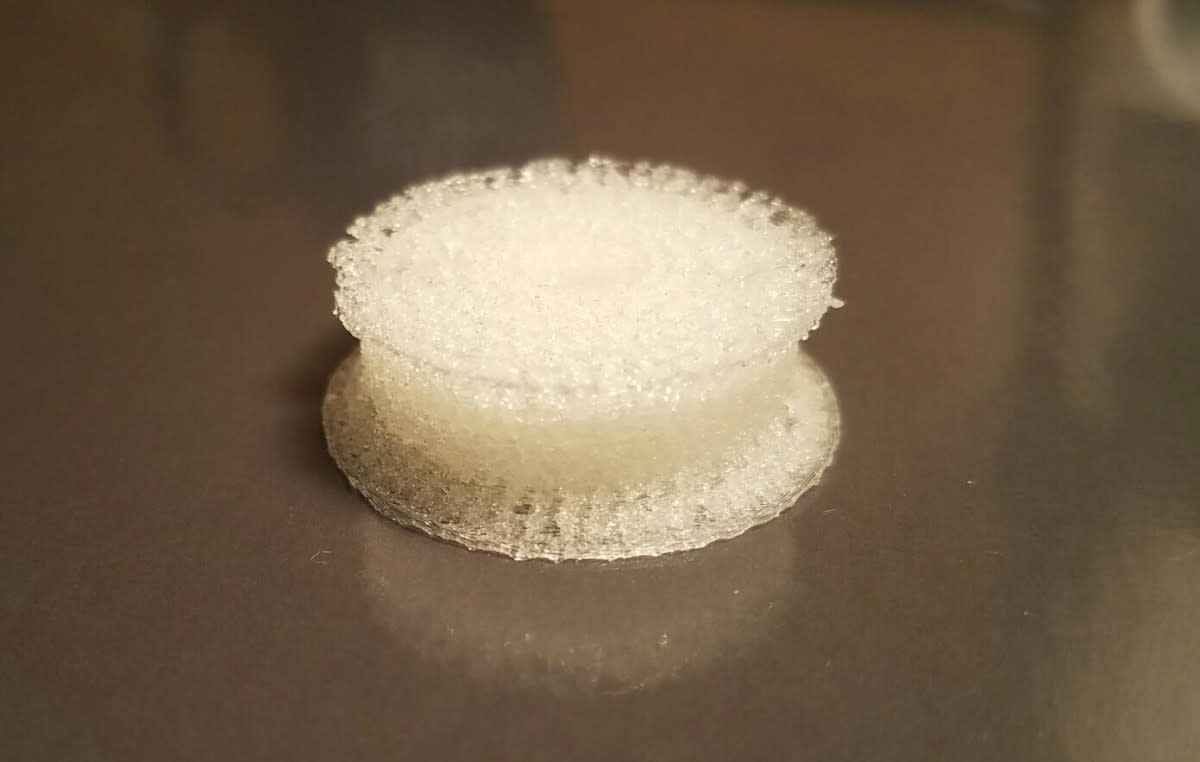
Eight cents.
That's all it costs to 3D-print a simple yet deceptively complex inch-and-a-quarter-wide plastic scaffold that could pave the way for surgeons to help patients regrow spinal bone tissue much more quickly and effectively.
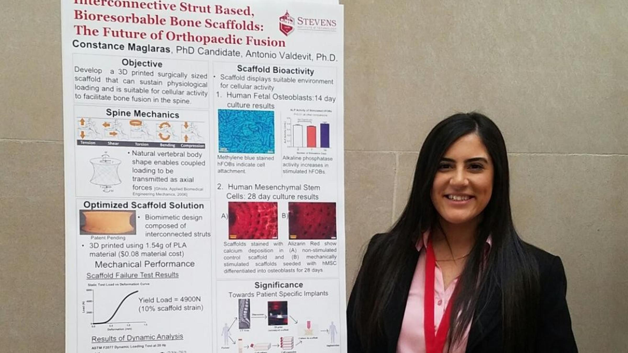
Stevens Institute of Technology biomedical engineering Ph.D. candidate Constance Maglaras, principal designer of the patent-pending technology, was recently awarded top prize in the graduate-poster division at Johnson & Johnson's 2017 Engineering Showcase for her poster and presentation, "Interconnective Strut Based, Bioresorbable Bone Scaffolds: The Future of Orthopaedic Fusion."
"This project is innovative, inspiring and full of hope for patients suffering from vertebral injuries. It brings together emerging technologies that will soon personalize healthcare in unimaginable ways," noted Johnson & Johnson Senior Director of Engineering and Design Jose Abreu. "We at Johnson & Johnson strive to improve the lives of all patients who use our products. This project aligns beautifully with our core principles."
Maglaras, who previously completed Stevens undergraduate and master's degrees, has worked since her sophomore year on innovations that aid spinal orthopedics. Thesis advisor Tony Valdevit, a biomedical engineering professor who has taught and supervised her research for seven years, is pleased with the work that culminated in her thesis and the prize-winning presentation.
"I knew her device would work well," says Valdevit. "We didn't know it would work this well."
Building a better scaffold for new vertebrae to grow on
Patients with spinal injuries preparing for spinal fusion surgery require implants. Traditional techniques require doctors to use autografts (bone removed from elsewhere in the patient, usually the pelvis), cadaver-sourced bone, or synthetic bone in those implants to aid in the fusion process.
New techniques explored by Maglaras could help enable the production of implants crafted of each patient's own mesenchymal stem cells (MSCs). The cells can be cultured for about one month outside the body on plastic scaffolds, where they begin to grow into osteoblasts (bone cells) until they resemble human bone.
However, it has proven challenging to optimize the shape of the artificial scaffold. Stem cells require nourishment from surrounding liquid media as they grow, so the scaffold must include adequate spacing to allow the life-giving brew to flow deep enough into the center of the device. The device must also be strong enough to withstand repeated compressions as a patient walks, twists, lifts and moves.
"It's a difficult mix to get right," notes Maglaras, who says she went through dozens of iterations before determining the best shape, which hinges on hundreds of connected, microscopic struts within the center of the device. "And getting a printer to also print this complex shape correctly was another challenge."
The proof was in the load testing.
"As soon as we saw it come off the printer, we looked at each other and knew this was 'it,' " Maglaras recalls.
"I thought it would be very good mechanically," adds Valdevit. "It turned out to exceed my expectations, both in terms of the strength under compression and the fatigue testing. She tested 5 million cycles at 20 hertz, increasing the load with each test. The piece compressed, but it didn't break."
Customizing implants to each patient's needs
"What makes this special is that it can withstand loads greater than the human spine normally needs to," notes Maglaras. "No other scaffold that I've come across is so lightweight and can withstand such loads. Once the cells have matured within the scaffold, it is almost like implanting a piece of customized bone, because it is made of the patient's own cells. Theoretically, there is no immunological risk in the process."
The scaffold can even be customized to an individual patient's physiology and bone-mineral density, she adds, by tweaking its design based on CT scans of a patient.
"That's personalizing the medicine here in the truest sense," she says.
After graduating in spring 2017, Maglaras is studying the possibility of seeking potential investors to create a startup based on the scaffold and attempting to commercialize or license the technology — "that would be very exciting," she says — while continuing to refine the design.
It's also possible she might enter the medical device industry instead, to work on further innovations that will benefit orthopedic practice. Valdevit has no doubt Maglaras will succeed on her chosen path.
"She has been a model student since day one," he says. "If she has a question, if she doesn't know something, she'll ask directly. And if she already knows the answer, if she knows what she needs to do, she is smart enough to just go and do it in the lab. I've also seen her teach and she's very, very good at that as well. We're very proud of her."