From 2D to 3D: A Newly Enhanced Imaging Technique Captures Brain Movement in Stunning Detail, Holds Potential to Inform Diagnosis of Brain Disorders
Researchers from the Mātai Medical Research Institute, in New Zealand, and Stevens Institute of Technology -- and others – report a new and enhanced method to vizualize difficult-to-spot brain conditions
(Hoboken, N.J. and Gisborne, New Zealand – May 5, 2021) – Magnetic Resonance Imaging (MRI) images are usually meant to be static. But now, researchers from Mātai Medical Research Institute (Mātai), Stevens Institute of Technology, Stanford University, the University of Auckland and other institutions, report on an imaging technique that captures the brain in motion in real time, in 3D and in stunning detail, providing a potential diagnostic tool for detecting difficult-to-spot conditions such as obstructive brain disorders and aneurysms – before they become life threatening.
The new technique, called 3D amplified MRI, or 3D aMRI, reveals pulsating brain movement which could help researchers to non-invasively visualise brain disorders and inform better treatment strategies for tiny deformations or disorders that obstruct the brain or block the flow of brain fluids.
Samantha Holdsworth, director of research at Mātai, senior lecturer at the University of Auckland and principle investigator at the Centre for Brain Research, and Mehmet Kurt, an assistant professor of mechanical engineering at Stevens Institute of Technology, have now published two papers on aMRI in collaboration with Stanford University, the University of San Diego California, Queens University, and the Icahn School of Medicine at Mount Sinai.
The first paper, published online today in Magnetic Resonance in Medicine, presents the 3D aMRI method, comparing it with its 2D aMRI predecessor. The new method results in a stunning visualization of the human brain’s movement that can be seen in all directions. The second paper, published online today as well in Brain Multiphysics, visualizes, validates and quantifies both the amplitude and direction of the brain as it moves in three dimensional space. The validation and quantification ensures that the software processing reflects an amplified version of real movement.
The approaches reported in the two papers could hold important clinical insights for a number of brain disorders. For example, the abnormal motion of two areas at the base of the brain, the pons and cerebellum, has been proposed as a diagnostic marker of Chiari I malformation, an abnormality that causes brain tissue to extend into the spinal canal.
2D amplified MRI was developed by Holdsworth, Mahdi Salmani Rahimi, Itamar Terem and other collaborators at Stanford University, enabling MRI imaging to capture brain motion in a way that had previously never been seen before. 3D amplified MRI builds on this previous work developed and published in 2016. The aMRI algorithm uses a video motion processing method developed by Neal Wadhwa, Michael Rubinstein, Fredo Durand, William Freeman and colleagues at Massachusetts Institute of Technology.
“The new method magnifies microscopic rhythmic pulsations of the brain as the heart beats to allow the visualization of minute piston-like movements, that are less than the width of a human hair,” explained Itamar Terem, a graduate student at Stanford and lead author of the first paper. “The new 3D version provides a larger magnification factor, which gives us better visibility of brain motion, and better accuracy.”
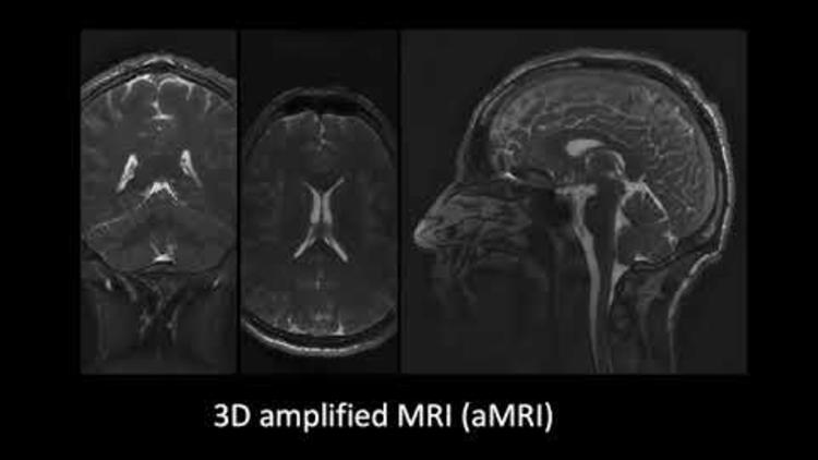
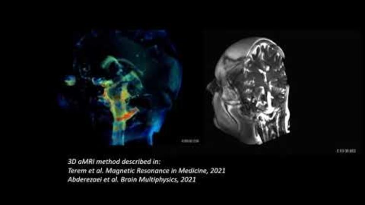
The 3D aMRI method, showing exquisite brain motion that is captured in all three planes of the brain (coronal, axial and sagittal views). Previously amplified motion was only reliably visible in the sagittal plane – the 3D aMRI method now captures motion in all planes. Outlined in Terem et al. Magnetic Resonance in Medicine (2021); Abderezaei et al. Brain Multi-physics (2021). https://youtu.be/bC05R_tcyW4
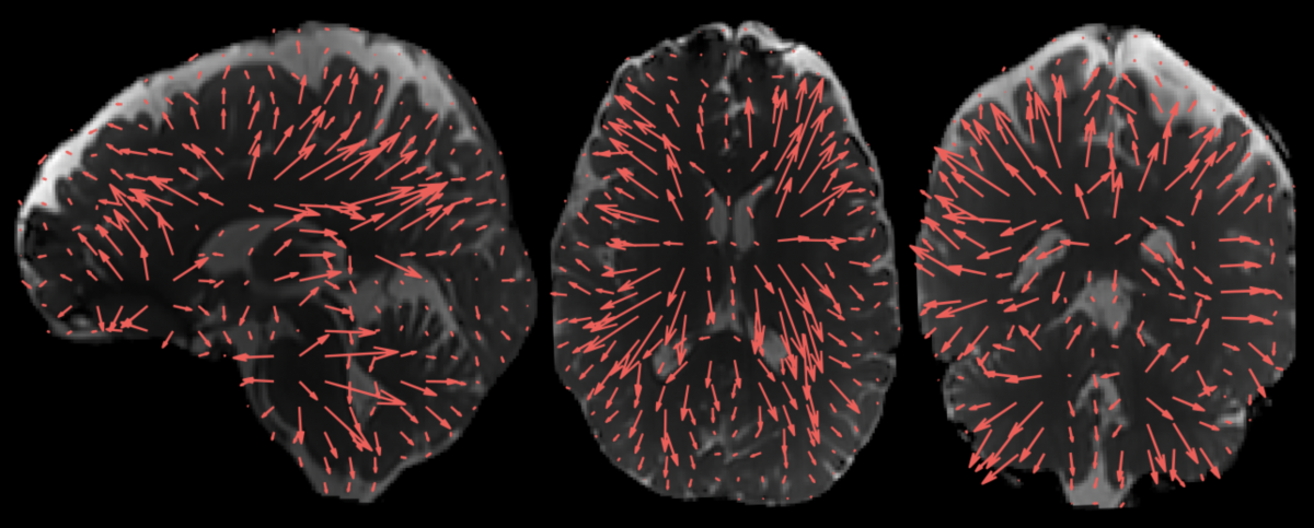
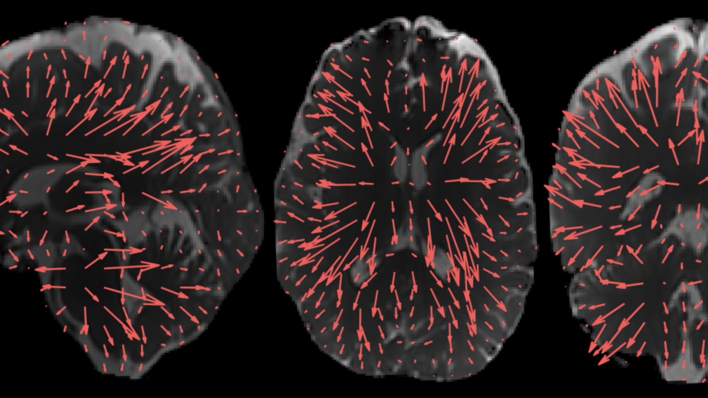
The arrows show the direction and amplitude of the brain’s movement. These displacement patterns, which were enabled by extra processing of 3D aMRI, may help us understand how the brain moves with different disorders. 3D aMRI method outlined in Abderezaei et al. Brain Multiphysics (2021); Terem et al. Magnetic Resonance in Medicine (2021).
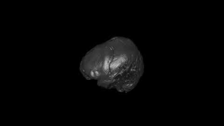
Using the new 3D aMRI software, 4D animation models of brain motion can be created from an MRI image. The striking detail of these animated magnified movements may be able to help identify abnormalities, such as those caused by blockages of spinal fluids, which include blood and CSF (spinal fluid in the brain). 3D aMRI method outlined in Terem et al. Magnetic Resonance in Medicine (2021); Abderezaei et al. Brain Multi-physics (2021). https://youtu.be/mRsnPqK4LCQ
Video: 3D aMRI not only provides a stunning look inside the "beating brain", but it can also measure this physiological motion in all directions. Here, the amplitude of brain motion is overlayed for each brain slice and orientation in 3D. 3D aMRI method outlined in Abderezaei et al. Brain Multiphysics (2021); Terem et al. Magnetic Resonance in Medicine (2021__). https://youtu.be/Yfplh32y5GY
3D aMRI of the human brain shows minute movements of the brain at an unprecedented spatial resolution of 1.2mm3, approximately the width of a human hair. The actual movements are amplified (made larger, up to 25 times) to allow clinicians and researchers to view the movements in detail. The striking detail of these animated magnified movements may be able to help identify abnormalities, such as those caused by blockages of spinal fluids, which include blood and cerebrospinal fluid.
“We showed that 3D aMRI can be used for the quantification of intrinsic brain motion in 3D, which implies that 3D aMRI holds great potential to be used as a clinical tool by radiologists and clinicians to complement decision making for the patient’s treatment,” said Mehmet Kurt, from the Stevens Insitute of Technology and senior author of the second paper. “In my lab at Stevens, we are already seeing the benefits of using variants of 3D aMRI technique in a variety of clinical conditions including Chiari Malformation I, hydrocephalus, and aneurysms, in collaboration with clinicians at Mount Sinai."
A number of research projects are underway using the new imaging software. Holdsworth said, “We are using 3D aMRI to see if we can find new insights into the effect of mild traumatic brain injury on the brain. She added, “One study already underway, a collaboration between Mātai and the University of Auckland, uses 3D aMRI together with brain modelling methods to see whether we can develop a non-invasive way of measuring brain pressure, which may in some cases remove the need for brain surgery”. This could be valuable clinically, for example, in children with idiopathic intracranial hypertension who often require invasive brain pressure monitoring.
Miriam Sadeng, an associate professor at the University of Auckland in the department of anatomy and medical imaging, who is a physician and is an author on both papers said, “This fascinating new visualisation method could help us understand what drives the flow of fluid in and around the brain. It will allow us to develop new models of how the brain functions, that will guide us in how to maintain brain health and restore it in disease or disorder.”
“Validating the method through computational modelling gave us further confidence about the potential impact of this work,” said Javid Abderezaei, a graduate student in Kurt’s lab at Stevens and lead author on the second paper. “What is exciting to see is that the dominant displacement patterns in the healthy brain qualitatively matched with the underlying physiology, which means that any changes in the physiological flow as a result of a brain disorder should be reflected in the displacements we measure.”
The capability to view the differences in brain motion could help us better understand a variety of brain disorders. In the future, the technology could be expanded to use in other health disorders throughout the body.
About Stevens Institute of Technology
Stevens Institute of Technology is a premier, private research university situated in Hoboken, New Jersey. Since our founding in 1870, technological innovation has been the hallmark of Stevens’ education and research. Within the university’s three schools and one college, 7,300 undergraduate and graduate students collaborate closely with faculty in an interdisciplinary, student-centric, entrepreneurial environment. Academic and research programs spanning business, computing, engineering, the arts and other disciplines actively advance the frontiers of science and leverage technology to confront our most pressing global challenges. As Stevens celebrates its 150th anniversary, the university continues to be consistently ranked among the nation’s leaders in career services, post-graduation salaries of alumni, and return on tuition investment.
About Mātai
Mātai is a non-profit medical imaging research and innovation centre, based in Gisborne, New Zealand. Our team aims to enhance the capabilities of Magnetic Resonance Imaging (MRI) through research and development into advanced medical imaging software and machine learning.
The team is led by Dr Samantha Holdsworth, Mātai Research Director, Senior Lecturer at the University of Auckland and Principle Investigator at the university’s Centre for Brain Research. Samantha is a leading expert in human brain imaging and a pioneer of super-fast, high resolution MRI methods and amplified MRI (aMRI), a new method of visualising brain motion). Her work has included the development of new MRI methodologies to help diagnose disease earlier. Mātai has a collaborative national and global network of 32 affiliate faculty advisors and 37 medical research, scientific, and engineering advisors. These include world leaders in their respective fields across various disciplines, and who are committed to collaborating across a range of medical research projects.
MRI is unsurpassed in its capabilities to transform our understanding of health. It is safe, non-invasive, and can reduce the need for unnecessary invasive surgeries. Advances in medical imaging, which include scan clarity and shortening scan time, have the potential to redefine the way we identify disease and disorders, enable earlier interventions and treatments, and new ways of managing health disorders.
The establishment of Mātai was made possible through seed funding by the Provincial Growth Fund, with additional support from Trust Tairāwhiti, the University of Auckland, the JN Williams Memorial Trust and the HB Williams Turanga Trust, Pultron Composites, the Dame Bronwen Holdsworth and Dr. Peter Holdsworth Trust and many others.
For inquiries, please contact:
Jeanette Lepper
[email protected]
+64273790170
www.matai.org.nz
Thania Benios
[email protected]
917-930-5988
Stevens.edu