Strong Vision: New Biomedical Engineering Chair Works to Diagnose, Treat Eye Diseases
Researcher focuses on quicker diagnoses, more effective drug delivery system for diabetes-related ocular disorders
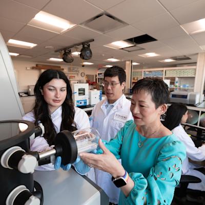
Incoming Stevens biomedical engineering department chair Jennifer Kang-Mieler has never met a challenge she didn't enjoy.
"I love learning new things, meeting new people, traveling new places, producing innovative research and new findings," she admits. "My own daughter says I can't sit still, and I'd say my daughter is probably 100% correct."
Now Kang-Mieler has taken on her most exciting professional challenge yet, trading Chicago's lake-effect snows and deep-dish pizzas for Hoboken's riverside views and easy access to the Big Apple as she works to take Stevens' signature biomedical engineering department to the next level.
"I'd like to make a little noise on campus, in a good way," she explains. "To get this department's story out there, let people know what we are doing so well to benefit society, and what we can do working with partners in the future."
Deep expertise in the human visual system
The child of parents who emigrated to the Midwest from South Korea when she was a teenager, Kang-Mieler studied mathematics, applied mathematics and — eventually — biomedical engineering at Northwestern University, where she found herself becoming progressively more interested in the clinical applications of engineering and design.
“Even as a mathematics major, I always enjoyed laboratory work,” she recalls. “ I found that I especially enjoyed biological research in the labs.”
 Working toward her Northwestern masters degree, Kang-Mieler joined a summer research project investigating the role of oxygen in eye function. That project cemented her interest in biomedicine — more specifically, on the diseases that affect our eyes and vision as we age.
"I was hooked," Kang-Mieler says, "on wanting to understand how the visual system works."
She would go on to spend 20 years as a respected faculty member at Illinois Institute of Technology in Chicago, developing growing expertise in ocular diseases, before joining Stevens this past June as a tenured faculty member and chair of the BME department.
Earlier diagnosis, better treatments for eye disease
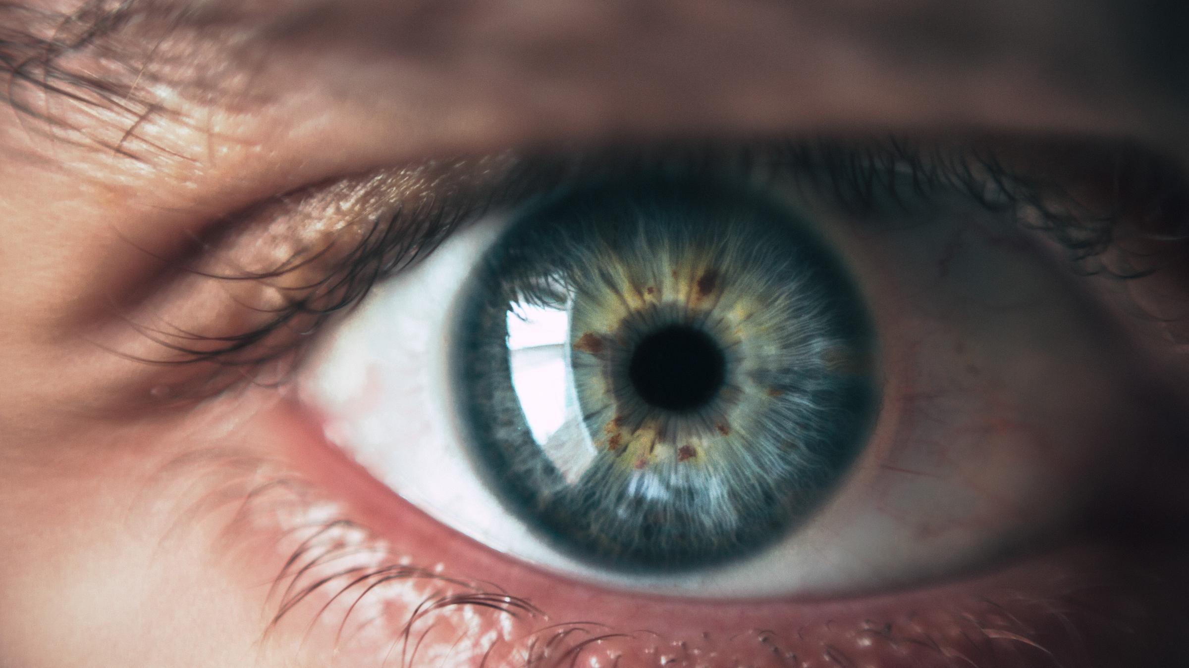
Kang-Mieler eventually developed specialized expertise in diabetic retinopathy (DR), a vision-loss disorder that eventually afflicts more than half of all diabetes patients
One area of her DR research involves developing earlier diagnosis tools for the disorder. The advanced form of the disease is caused when blood vessels begin growing too quickly in the eyes, eventually leaking, bursting or tugging on the retina, all of which reduce vision. But it can take time for clinical signs to show up.
"Even after being diagnosed with diabetes," she explains, "it takes six to seven years for DR symptoms to show. This is largely a silent disease for those first six years; the retina looks completely normal during that time, even with our best diagnostic imaging tools."
When DR is diagnosed earlier in its course, patients can change their diet, exercise and therapeutic regimens and slow or forestall progression of the disease, preserving vision for longer periods of time.
Diagnosing DR early, however, has proven a difficult challenge for clinicians.
"We have really good imaging tools and technologies to look at the eye's blood vessels, yet we still can only catch DR in its mid-stages — not usually its very early stages, when the changes and signs of leakage are more subtle."
So Kang-Mieler hit upon the idea of improving fluorescein angiography — a well-studied technique based on injection of dyes into the eye's blood vessels — by combining and processing image data with new algorithms in order to detect the telltale deterioration of blood vessels earlier in the disease's progression.
"This could become a very sensitive biomarker," she says of her technique, which leverages a so-called dynamic tracer kinetic model.
In 2021, the National Institutes of Health (NIH) agreed, awarding Kang-Mieler's work to enhance the novel technique with a $2.5 million award via the National Eye Institute. That support will run through 2026.
A second branch of her DR research works to develop improved delivery of medications directly to the eye's blood vessels to treat the disease and other eye disorders. Many ocular disorders are caused by blood vessels' unruly growth, which is spurred by overproduction of a human growth-spurring protein known as VEGF (vascular endothelial growth factor).
Anti-VEGF medications have recently emerged to suppress these growth spurts. But delivering those medicines precisely and successfully requires monthly injections directly into the eye, often for a lifetime, since protein-based antibody therapies quickly become washed out of, or inactive, in the body.
"Monthly injections for life: that's asking a lot of your patients," says Kang-Mieler. "Now we have developed a system that may work as well, or better, while requiring only one injection every six months."
To help the ocular medications persist longer, her team worked with material scientists to design patented microspheres embedded within heat-sensitive hydrogels. Therapeutic antibodies, injected within a liquid, quickly become suspended in a gel once they enter the eye — allowing them to stay in place, and active, far longer than they do with conventional injections.
Kang-Mieler was awarded a separate $2 million award from the NIH to support development of this proposed drug-delivery system, which has already proven successful in early simulations and small-animal models. (Primate research is the next and final step to complete successful before potentially receiving FDA approval for human trials.)
"It has been a long road to this point with these research projects," she says. "And I'm very excited to begin the next steps at Stevens."
Taking BME to the next level
Kang-Mieler also looks forward to leading an academic and research department that continues to grow in size and impact.
"The biomedical engineering faculty at Stevens are exciting," she says. "There is a lot of energy, youth, a lot of good ideas."
She already has ideas about new directions the department may take, and ways in which to communicate its work.
"We need to develop and tell our unique story about what this department does, working so closely with the medical field, helping people. We need to talk about how we are helping people with diabetes, developing new medical devices and new materials, helping people cope with aging," explains Kang-Mieler.
She will work, she says, to forge new ties with major medical centers and medical schools in the metropolitan New York region.
"A clinical partner can really accelerate getting things developed and to market," says Kang-Mieler. "I've found that direct access to clinicians is very important as you design and test new devices and technologies."
Along those lines, she suggests development of new medical-school curricular tracks for Stevens' most talented biomedical engineering majors.
"Our undergraduate honors students, for example, could plot a course that takes six to seven years to attain an M.D. at a companion medical school in some form of a joint program," she says.
Kang-Mieler would also like to develop new curricular offerings to better educate graduate biomedical engineering students on the design process, working in tandem with industry and medical experts.
"As engineers, we sometimes have a tendency to believe we have the ability to solve any problem simply by designing the perfect device or solution," Kang-Mieler notes. "But it's important to work with patients and clinicians while you are designing things, throughout that process, in order to learn whether it will really benefit patients in the end or not."
"You don't want to just design a fancy thing without any clinical utility," she concludes. "You need to explore all the possibilities, with the patient always at the center of the conversation."