Department of Biomedical Engineering
Academic Programs
Catalyze Your Career
Announcements
Hongjun Wang Appointed George Meade Bond Professor Through 2030
The Charles V. Schaefer, Jr. School of Engineering and Science is proud to announce the appointment of Dr. Hongjun Wang as the George Meade Bond Professor in Biomedical Engineering, effective July 1, 2025 through Aug. 31, 2030.
Wang joined Stevens in 2005 and has since advanced from assistant professor to full professor. He served as the founding chair of the Department of Biomedical Engineering (2018–2022) and most recently as director of the Semcer Center for Healthcare Innovation (2022–2025). A globally recognized scholar, Wang’s research spans biomimetic materials, multiscale tissue reconstruction, nanomedicine and regenerative medicine. He has secured over $6 million in external funding and ranks in the top 0.45% globally in the field of “scaffolding,” according to ScholarGPS.
A Fellow of the National Academy of Inventors, Dr. Wang is the recipient of numerous honors, including the New Jersey Innovators Award, the Provost’s Award for Academic Entrepreneurship & Enterprise Development, and the Jess N. Davis Award for Excellent Research. He holds five patents and has authored nearly 140 peer-reviewed journal articles, 12 book chapters and two edited volumes. His work has been widely presented through more than 100 invited talks, including keynote and plenary lectures.
This prestigious appointment recognizes Wang’s outstanding scholarship, teaching excellence and lasting contributions to Stevens and the biomedical engineering field.

CardioLink Project by Stevens Biomedical Engineering Graduates Wins NIH National Prize
The Department of Biomedical Engineering is proud to share that CardioLink — a project developed by recent Stevens graduates that won first place in the Ansary Entrepreneurship Competition at the 2025 Stevens Innovation Expo — has also been awarded the National Institute on Aging Prize in the NIH/VentureWell DEBUT Challenge.
CardioLink addresses a critical need in post-discharge care for heart failure patients, a group at highest risk for preventable readmissions. The tabletop device simplifies monitoring by combining weight, blood pressure, heart rate, oxygen saturation, medication adherence, and daily behaviors into one five-minute daily check-in. Unlike mobile apps or invasive systems, CardioLink reduces patient burden while delivering meaningful insights to clinicians.
Central to the device is a proprietary Multi-Factor Health Index (MFHI), which condenses multiple health indicators into a single risk score. This innovation enables earlier intervention, helping clinicians reduce readmissions and improve outcomes.
The team — biomedical engineering alumni Robert Gordanier, Rhys Robichaud and Panos Stamas — developed the system under faculty and clinical mentorship. Their work has already drawn interest from major hospital networks, and intellectual property protection has been filed to support commercialization. The team will present their research at the Biomedical Engineering Society Annual Conference in San Diego this October.
We congratulate the CardioLink team for this exciting advancement in health innovation with the potential for real-world clinical impact.

Stevens Biomedical Engineering Graduates Win National Prize for Breast Health Innovation
The Department of Biomedical Engineering is pleased to announce that DensiSense, a senior design project developed by recent Stevens graduates, has earned national recognition in the 2025 DEBUT Challenge. Sponsored by the National Cancer Institute, the competition drew 123 applications from 67 universities across 24 states. The Stevens team received the Technologies for Cancer Prevention, Diagnosis, and Treatment Prize and a $15,000 award.
DensiSense is an at-home, wearable breast health monitoring system designed for women with dense breast tissue, a population at greater risk for breast cancer. While these patients are often recommended for more frequent screenings, long intervals between appointments and the limitations of traditional self-exams leave many without effective options to track their health.
The system integrates a smart sports bra with a specialized sensor capable of measuring breast tissue stiffness. These measurements transform self-exams from subjective guesswork into reliable, trackable data. Results are analyzed with artificial intelligence to determine whether they fall within healthy ranges or call for medical evaluation. The goal is to provide women greater control over their breast health through technology that fits into everyday life, closing the gap between routine exams and real-world monitoring for more timely information and improved peace of mind.
The project was led by recent biomedical engineering graduates Sonali Dalwadi and Simran Salem, who filed a provisional patent during their senior year and are working toward a full patent in 2026. They will present their work at the Biomedical Engineering Society Annual Conference in San Diego this October.
The department is proud of our students’ accomplishment and excited to follow their next steps in advancing this promising technology.

Dr. Jennifer Kang-Mieler Appointed Director of Semcer Center for Healthcare Innovation
The Charles V. Schaefer, Jr. School of Engineering and Science is pleased to announce the appointment of Dr. Jennifer Kang-Mieler as director of the Semcer Center for Healthcare Innovation (CHI), which became effective on July 1, 2025.
Dr. Kang-Mieler, George Meade Bond Professor and Chair of the Department of Biomedical Engineering, joined Stevens in 2022. A nationally and internationally recognized leader in ophthalmology and vision science, her research focuses on retinal vascular diseases and the development of technologies that improve clinical diagnostics and treatment. Her work is currently supported by three active NIH R01 grants and has received funding from the NIH, the Whitaker Foundation, the Lincy Foundation, the Macula Foundation and industry collaborators.
As director, Dr. Kang-Mieler will lead efforts to expand CHI’s research and educational initiatives, foster interdisciplinary collaboration and build strategic partnerships with clinical and industry stakeholders. Her leadership will be instrumental in advancing CHI’s mission to drive innovation in healthcare technology.
We also extend our sincere gratitude to Dr. Hongjun Wang for his outstanding leadership over the past three years. Under his direction, CHI was relaunched with a renewed vision, strengthened research culture and new collaborations with regional medical institutions.

Hongjun Wang Appointed George Meade Bond Professor Through 2030
The Charles V. Schaefer, Jr. School of Engineering and Science is proud to announce the appointment of Dr. Hongjun Wang as the George Meade Bond Professor in Biomedical Engineering, effective July 1, 2025 through Aug. 31, 2030.
Wang joined Stevens in 2005 and has since advanced from assistant professor to full professor. He served as the founding chair of the Department of Biomedical Engineering (2018–2022) and most recently as director of the Semcer Center for Healthcare Innovation (2022–2025). A globally recognized scholar, Wang’s research spans biomimetic materials, multiscale tissue reconstruction, nanomedicine and regenerative medicine. He has secured over $6 million in external funding and ranks in the top 0.45% globally in the field of “scaffolding,” according to ScholarGPS.
A Fellow of the National Academy of Inventors, Dr. Wang is the recipient of numerous honors, including the New Jersey Innovators Award, the Provost’s Award for Academic Entrepreneurship & Enterprise Development, and the Jess N. Davis Award for Excellent Research. He holds five patents and has authored nearly 140 peer-reviewed journal articles, 12 book chapters and two edited volumes. His work has been widely presented through more than 100 invited talks, including keynote and plenary lectures.
This prestigious appointment recognizes Wang’s outstanding scholarship, teaching excellence and lasting contributions to Stevens and the biomedical engineering field.
CardioLink Project by Stevens Biomedical Engineering Graduates Wins NIH National Prize
The Department of Biomedical Engineering is proud to share that CardioLink — a project developed by recent Stevens graduates that won first place in the Ansary Entrepreneurship Competition at the 2025 Stevens Innovation Expo — has also been awarded the National Institute on Aging Prize in the NIH/VentureWell DEBUT Challenge.
CardioLink addresses a critical need in post-discharge care for heart failure patients, a group at highest risk for preventable readmissions. The tabletop device simplifies monitoring by combining weight, blood pressure, heart rate, oxygen saturation, medication adherence, and daily behaviors into one five-minute daily check-in. Unlike mobile apps or invasive systems, CardioLink reduces patient burden while delivering meaningful insights to clinicians.
Central to the device is a proprietary Multi-Factor Health Index (MFHI), which condenses multiple health indicators into a single risk score. This innovation enables earlier intervention, helping clinicians reduce readmissions and improve outcomes.
The team — biomedical engineering alumni Robert Gordanier, Rhys Robichaud and Panos Stamas — developed the system under faculty and clinical mentorship. Their work has already drawn interest from major hospital networks, and intellectual property protection has been filed to support commercialization. The team will present their research at the Biomedical Engineering Society Annual Conference in San Diego this October.
We congratulate the CardioLink team for this exciting advancement in health innovation with the potential for real-world clinical impact.
Department Facts
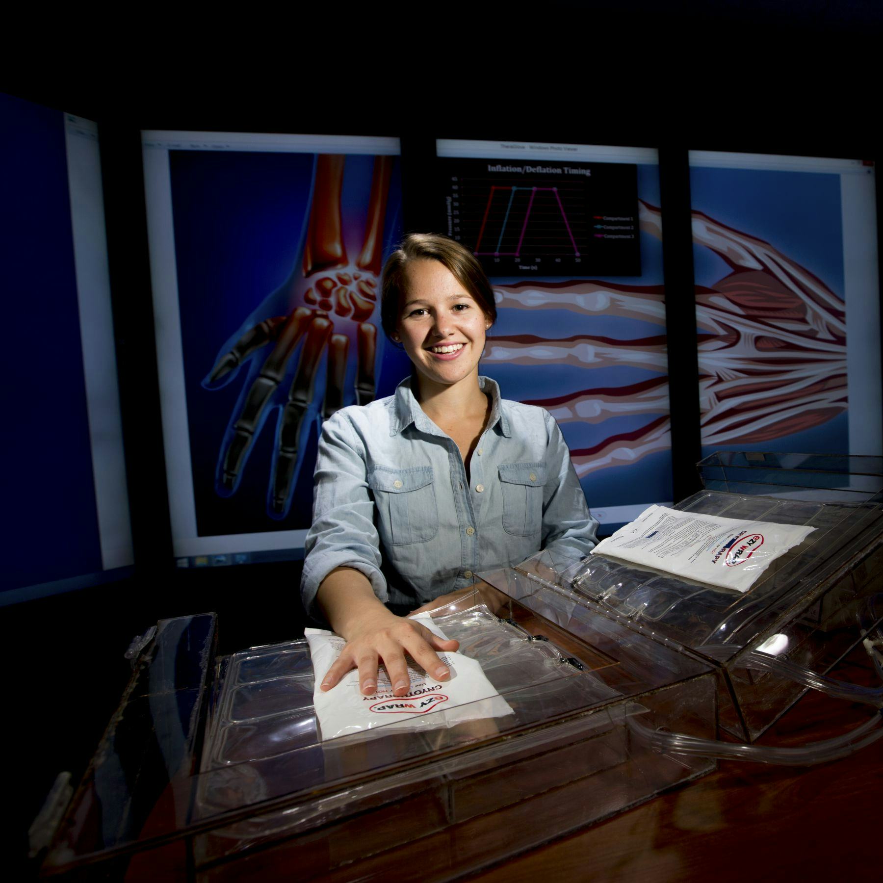
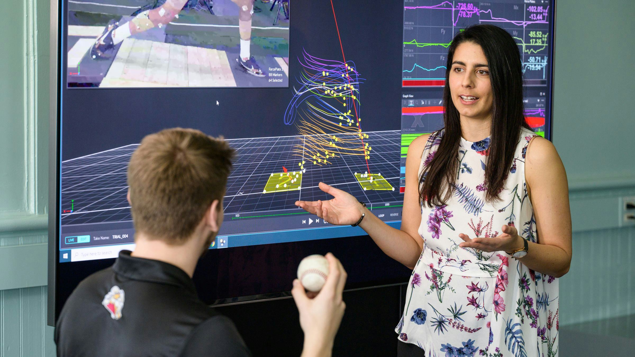
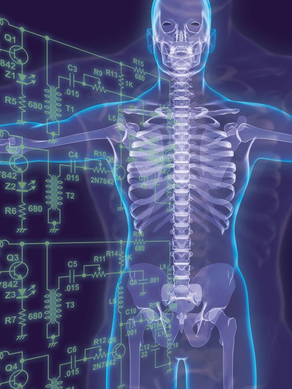
Biomedical Engineering Research
Our entrepreneurial environment encourages technological innovation from concept to commercialization with a focus on advancing biomedical technology, healthcare delivery and nanotech applications.
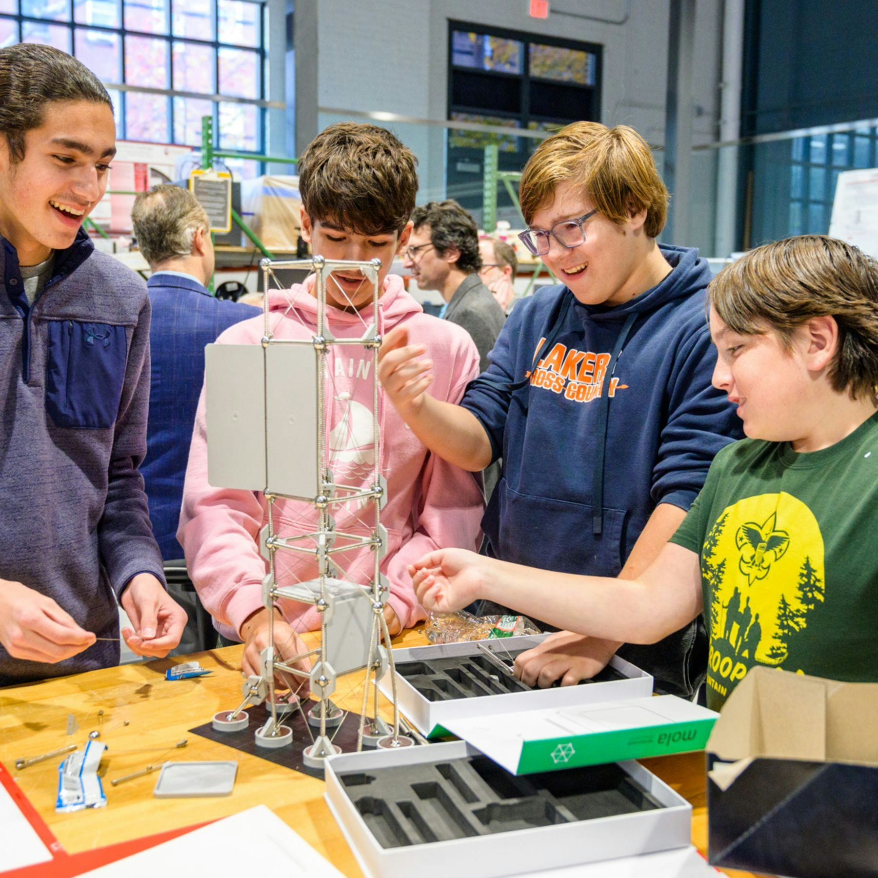
STEM Outreach at the Schaefer School
The Charles V. Schaefer, Jr. School of Engineering and Science is dedicated to science, technology, engineering, and math education and enrichment at all levels. Our numerous STEM outreach and education programs are designed to instill a love of science and technology in both teachers and students from K - 12 throughout the New York and New Jersey area, and beyond.
Get Social With Us
What’s happening in the Department of Biomedical Engineering at Stevens? Follow us on Instagram and LinkedIn for student life, faculty highlights and research in action.
Department News


Upcoming Seminars and Events


Department Leadership
Meet the faculty and staff leading the Department of Biomedical Engineering. Find contact information for department administrators, academic advisors and more.
