Stevens Researcher and RadioSight Partner Awarded $256,000 NSF Grant for Handheld Skin Cancer Imaging Device
The Small Business Technology Transfer grant will help Negar Tavassolian and Amir Mirbeik ’18 develop a handheld device that helps diagnose skin cancer and guide surgical decisions
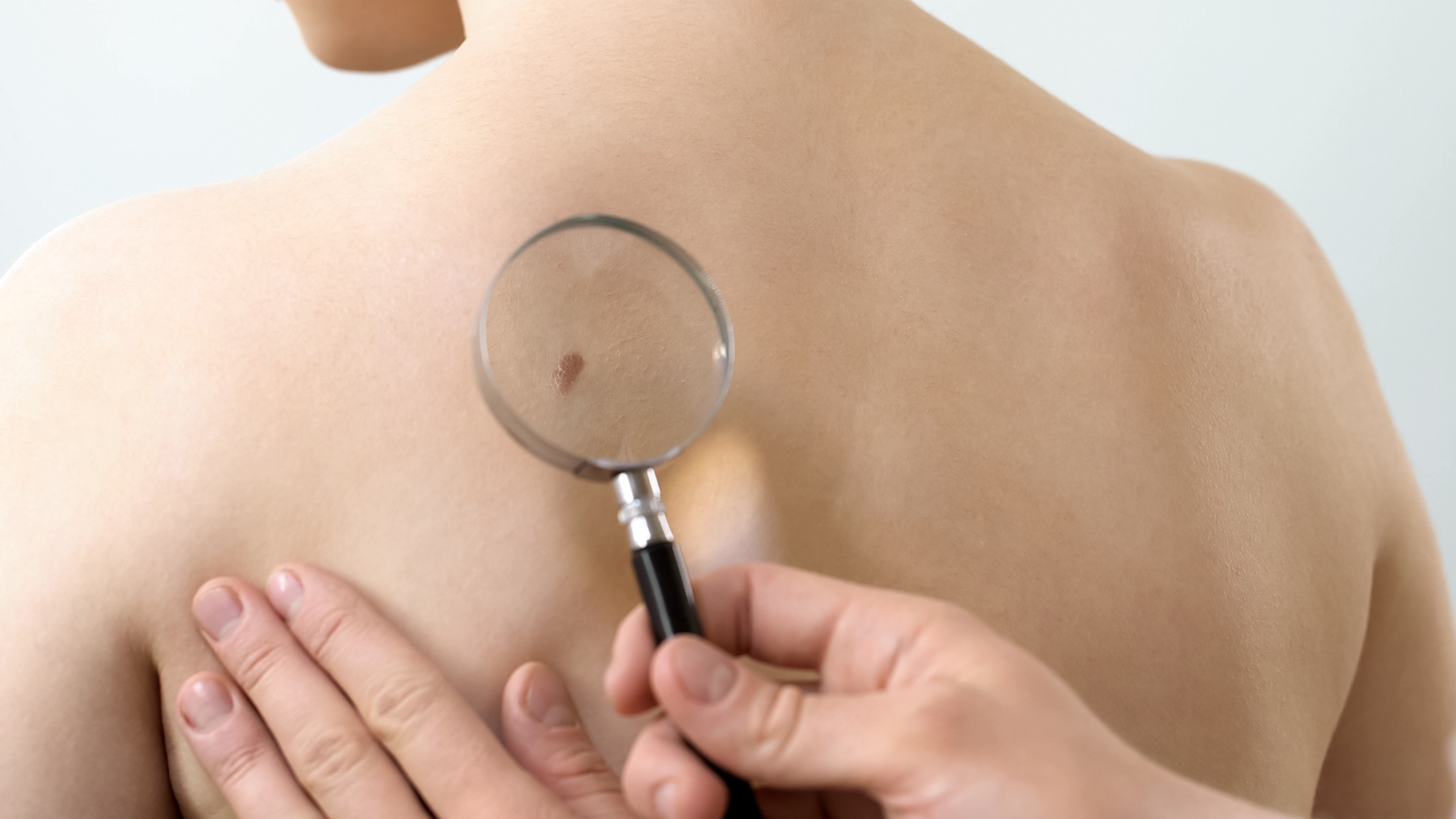
In the United States alone, two people die from skin cancer every single hour. It’s the most common cancer around the world — and a researcher at Stevens Institute of Technology is coming for it.
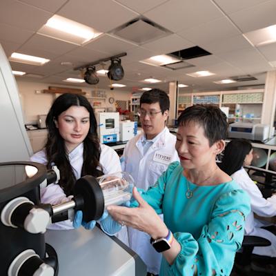
The National Science Foundation (NSF) recently awarded a $256,000 Phase I Small Business Technology Transfer grant to associate professor of electrical and computer engineering Negar Tavassolian and her RadioSight partner and former doctoral fellow Amir Mirbeik ’18, of which $80,000 directly funds research at Stevens. The grant supports the development of a novel handheld device that dermatologists and dermatologic surgeons can use when diagnosing and treating skin cancer.
Putting Cancer Detection Support in Doctors’ Hands
Right now, there simply isn’t tech available to help dermatologists visualize tissues when diagnosing skin cancer or making surgical decisions during biopsy.
“Surgeons just use their eyes to remove the tissue,” explained Tavassolian. “There's no imaging modality to help them picture it, so a lot of times some unnecessary tissue is removed because of lack of accuracy. And sometimes [cancerous] tissues are not completely removed, and people have to go back and do another biopsy.”
Tavassolian envisions a future where every dermatologist’s office has a handheld 3D imaging device that shows the underlying shape and profile of suspicious tissue, helping doctors differentiate between normal tissue and tissue that requires biopsy. A dermatologic surgeon would use the device to visualize the margins before biopsy to improve accuracy when excising skin lesions.
Tavassolian and Mirbeik have already secured a US patent and another provisional patent for different aspects of their technology. The one-year NSF grant will support phase I development, proving the efficacy of the team’s prototype and eventually scaling it into a handheld tool.
At this point, that prototype is pretty big—about the size of a series of small tables. The team already installed the prototype in a dermatologist’s office for an in vivo study in collaboration with Hackensack University. There, the device was used during medical appointments with 120 dermatology patients to demonstrate its utility in distinguishing between skin cancer, benign lesions, and normal tissues. The results are now under review in a scientific journal.
Enhanced Imaging for Medical Applications
To produce those 3D views, the device relies on a form of millimeter wave imaging. That’s the technology used by body scanners in airports — but Tavassolian and Mirbeik are amping up and fine tuning its capabilities.
“Millimeter wave imaging that’s available right now is used for commercial applications and also military applications due to limited bandwidth and limited resolution in the images that it provides,” said Tavassolian. “In our research, we worked on improving the resolution by improving the bandwidth. So that's how we got the ultra-wideband millimeter wave imaging. And once we improve the resolution, if we have ultra-wideband, we can use it for medical applications—for example, skin cancer detection.”
The research team will use the integrated circuit (IC) technology—the same tech that makes it possible for a cellphone to work like a computer yet fit in your palm—to scale the product into a handheld device about the size of a camera. The ultimate goal is for the device to be an accessibly-priced staple in every dermatology practice.
Eventually, Tavassolian hopes to enhance the device by enabling it to provide pop-up recommendations to help guide clinicians. Plus, since the technology acts like a 3D magnifying glass, highlighting the contrast between tumors and normal tissues, it’s possible it could even work for other high-contrast medical applications — like detecting tooth decay or monitoring things like wound healing or blood glucose levels.
For Tavassolian, whose studies and career have brought her from Iran to Canada and then to the United States, the best part of her work is that it opens new doors.
“I really enjoy research and moving in new directions and learning new things and doing things that others haven't done,” she explained. “When a paper comes out, I'm really excited. When we get new results, I'm really excited. All stages are really exciting for me.”
For the 9,500 people who will receive a skin cancer diagnosis today, Tavassolian’s vision could mean a more hopeful future.
Learn more about electrical and computer engineering at Stevens:
Department of Electrical & Computer Engineering
Learn more about research in the Department of Electrical and Computer Engineering →